


The microscopy lab enhances the quality of imaging to extremely high.
11. 8. 2022
The Department of Advanced Materials at CXI shows how important it is to upgrade cutting-edge instrumentation. It can now record nanostructures at extremely high resolution.
Researchers in the Microscopy Laboratory have shown that innovation can also be applied to the refurbishment of technical equipment. A scanning electron microscope (SEM) is an optical instrument that can image the structures of samples at extremely high resolution. The resolution of the UHR-SEM is around 1 nm.
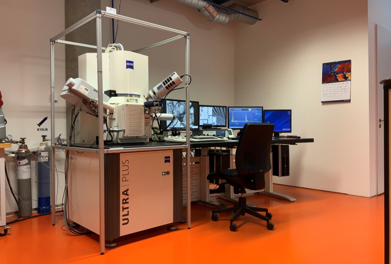 Zeiss Ultra Plus scanning electron microscope with Spicer SC24 cage installed to compensate for electromagnetic interference.
„Just simply: we are increasing the resolution. The electron microscope's parameters far exceed the capabilities of optical microscopes. While the resolving power of the human eye is approximately 0.2 mm, the thickness of two office papers, that of an optical microscope is about 0.2 µm, a thousand times higher. With an electron microscope, we are getting to a resolution of 1 nm," says Pavel Kejzlar, a researcher in the microscopy team.
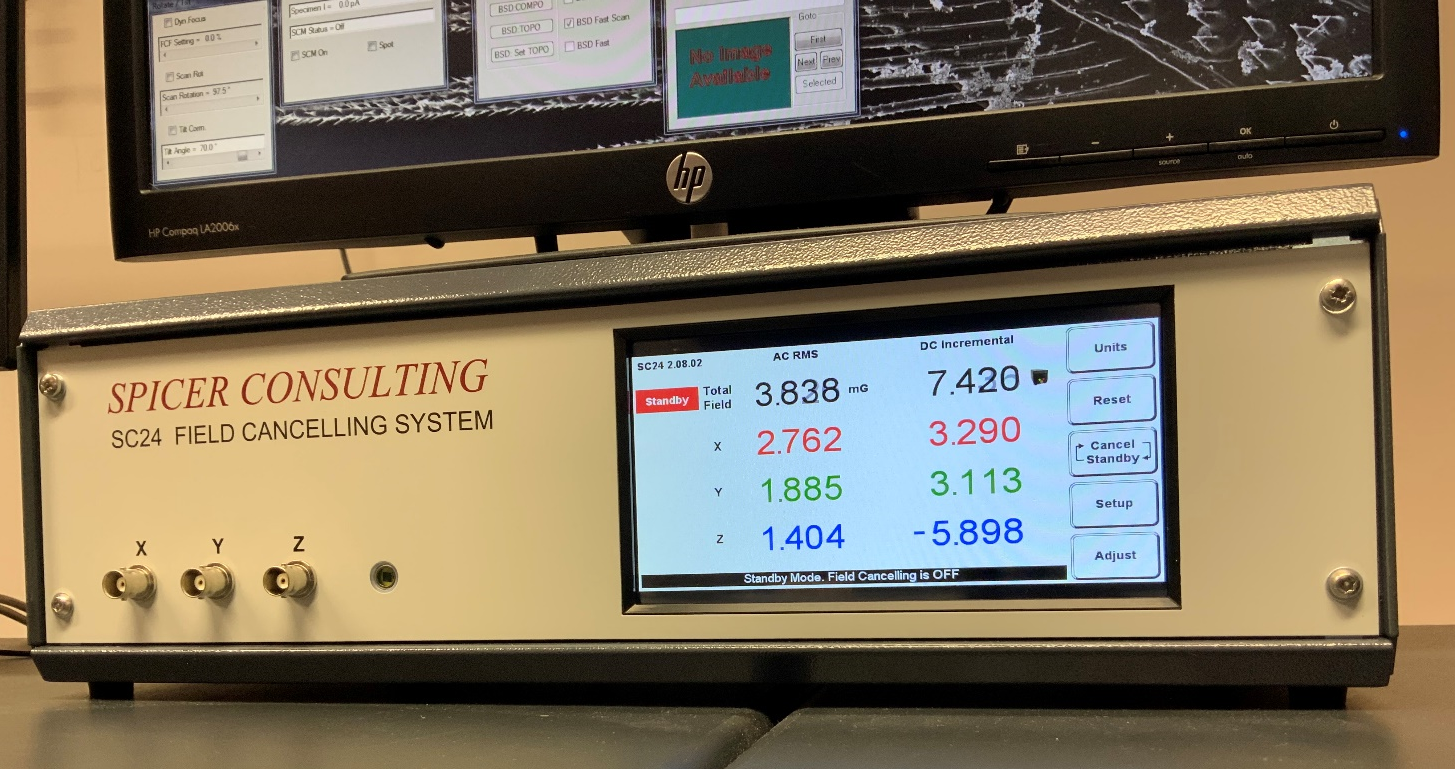
Just for comparison: the values on the monitor are measured before the new system was activated.
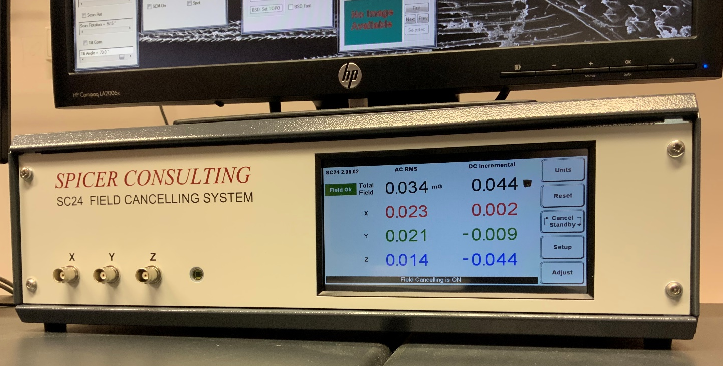
By activating the newly purchased system, it is possible to achieve more than 100-fold reduction in the intensity of AC and DC interference. The figure shows the measured values after activation.
Achieving such measurement accuracy means meeting conditions for minimal vibration and noise, temperature stability and ensuring the absence of electromagnetic interference. Its interfering signal undesirably affects the function of electronic devices. This signal is usually generated by the surrounding electrical equipment and the power grid. The primary electron beam reacts to changes in the electromagnetic field by deflecting, resulting in loss of resolution and undesirable image distortion.
Therefore, it was necessary to purchase a system to compensate for the disturbing electromagnetic field, the SPICER SC24. The SC24 stabilizes the field by dynamically creating an almost equal but opposite field, which makes the microscope's imaging performance significantly better, describes Mateusz Fijalkowski, head of the microscopy lab.
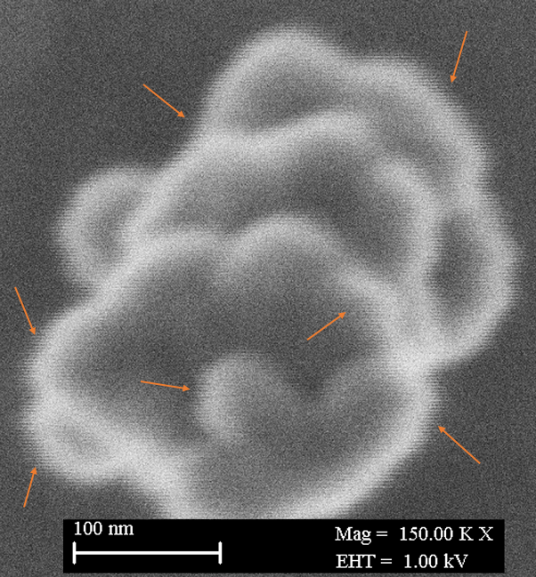
Beam smearing in line scanning is clearly visible especially at the edges of the observed sample when higher magnifications and low accelerating voltages are used, when the primary electron beam is more susceptible to ambient interference (here power grid interference at 50Hz).
The SC24 system is controlled by a control unit, equipped with a magnetic field sensor and three orthogonal axial multicore cables that are installed in a frame designed around the microscope. The magnetic field cancellation method is based on broadband analog negative feedback. A magnetic field sensor measures the intensity of magnetic field changes in real time. A microcomputer embedded in the control unit digitises the measured field and then controls three power amplifiers. These generate currents in the cables to produce an oppositely polarised field of the same magnitude. The whole system can dynamically react to field changes and compensates automatically within 100 µs.
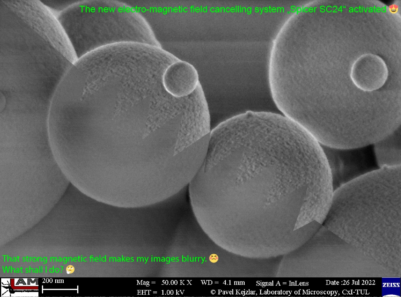
The electromagnetic field oscillates the primary electron beam, resulting in image blurring and loss of important information about the sample structure.
.png)
.jpeg)
.png)
.jpeg)
.png)
.jpeg)
.png)


.png)